
Sub-Millimeter Cuvettes (0.01-0.5 mm): When 1 mm Is Too Long
This post is also available in:
A sub-millimeter cuvette is a quartz cell with path length below 1 mm (typically 0.01, 0.05, 0.1, 0.2, or 0.5 mm), designed for high-absorbance samples where a standard 10 mm cell would saturate the detector. Concentrated proteins (≥ 50 mg/mL), undiluted dyes, neat solvents in NIR, and high-OD pharma intermediates all require sub-mm cells to bring absorbance into the linear Beer-Lambert range of 0.1–1.0 AU.
Sub-Millimeter Path · High-Absorbance Samples
Sub-Millimeter Cuvettes (0.01–0.5 mm): When 1 mm Is Too Long
Concentrated proteins above 50 mg/mL, plasmid DNA above 1,000 ng/µL, neat oils, undiluted dyes, and crude reaction mixtures saturate 1 mm cuvettes and even most NanoDrop pedestals. Sub-millimetre quartz cells (0.01, 0.05, 0.1, 0.2, 0.5 mm) and demountable thin-film holders bring these samples back into the linear absorbance window without the dilution step that costs sample, time, and accuracy.
0.01–0.5 mm
Sub-mm Path Range
100x
vs 10 mm reduction
5–100 µL
Sample Volume
No Dilution
Direct Measurement
The standard 10 mm cuvette is the workhorse of UV-Vis spectroscopy because most samples sit comfortably in its 0.1–1.0 AU detection window at typical lab concentrations. But “typical” excludes a meaningful fraction of analytical work: undiluted recombinant antibody at 50–200 mg/mL, plasmid prep at 2–3 µg/µL, neat fuel oils, undiluted reaction-mixture dyes, glycerol-stabilised DNA at μg/µL concentrations. In a 10 mm cell every one of these saturates the spectrophotometer at well above 2.0 AU. The standard fix — serial dilution — introduces pipetting error, contamination risk, and consumes sample.
The mechanical fix is to shorten the path length. A 0.1 mm cuvette gives you 100x less absorbance than a 10 mm cell at the same concentration, bringing the same neat sample back into the 0.1–1.0 AU window without dilution. Sub-millimetre cells (0.01, 0.05, 0.1, 0.2, 0.5 mm) and demountable thin-film holders are how laboratories measure concentrated samples directly. This guide covers when to reach for them, how to choose between path lengths, the filling technique that gets reliable readings (sub-mm cells are unforgiving of bubbles), and the SKUs MachinedQuartz keeps in stock.
Sub-mm vs NanoDrop pedestal. Microvolume pedestals (NanoDrop One, Implen NanoPhotometer) auto-shorten the optical path to about 0.2 mm for high-concentration samples and reach 1–2 µL sample volumes. For sample-volume-limited DNA QC, the pedestal is the right tool. For everything else where path length below 1 mm matters, a sub-mm quartz cuvette is more accurate, more traceable, more flexible, and 50x cheaper than buying a pedestal. See the cuvette vs NanoDrop comparison for the full discussion.
1. Why standard cuvettes fail at high concentration
Modern UV-Vis spectrophotometers saturate at approximately 2.0–3.0 AU depending on instrument design. Above the saturation threshold, the detector cannot distinguish the small amount of transmitted light from background. The Beer-Lambert law — A = ε · c · l — tells you that absorbance scales linearly with concentration, so a sample that reads 0.5 AU at 1 mg/mL in a 10 mm cell will read 50 AU at 100 mg/mL. The instrument can’t measure that; either you dilute or you reduce path length.
Why dilution is the wrong answer for many samples
- Pipetting error compounds. A 1:1000 dilution typically requires three serial 1:10 dilutions. Each 1:10 step has a 1–3 % systematic pipetting error. Three steps stack to 5–10 % cumulative error before you even read the spectrometer.
- Sample is consumed. A 1 mg/mL final read from a 100 mg/mL stock requires preparing 100 µL of dilute material from 1 µL of stock — or larger volumes for larger reads. Precious-sample workflows (single-cell proteomics, gene therapy vectors, primary biological extracts) cannot afford the loss.
- Diluent contamination risk. Buffer-dependent assays (where the diluent contributes to the spectrum) require analytically clean buffer matched to the stock; impurities concentrate in the dilution.
- Speed. A 1:1000 dilution takes 5–10 minutes per sample. A direct measurement in a 0.1 mm cell takes 30 seconds.
For samples where any of these matter, the right answer is to keep the original concentration and reduce path length.
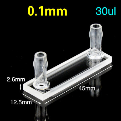 0.1 mm flow cell
C012WS5 — 0.1 mm flow cuvette
30 µL · molded 83 · for high-throughput concentrated DNA & protein QC
View C012WS5 → 0.1 mm demountable
C0.12WE — 0.1 mm detachable
30 µL · disassembles for cleaning · for viscous and particulate samples
View C0.12WE → 0.5 mm sealed
C0.54TE — 0.5 mm screw-cap
175 µL · four-way light · screw cap for kinetics, volatile or anaerobic samples
View C0.54TE →2. Choosing between sub-millimetre path lengths
The available standard sizes from MachinedQuartz are 0.5, 0.2, 0.1, 0.05, and 0.01 mm. The choice depends on how concentrated your sample is and what absorbance you target.
| Path length | Reduction vs 10 mm | Best concentration range (approx.) | Typical sample volume | Format |
|---|---|---|---|---|
| 0.5 mm | 20x | 10–30 mg/mL protein; 200–1000 ng/µL dsDNA | ~ 100 µL | Standard sub-micro cuvette |
| 0.2 mm | 50x | 20–80 mg/mL protein; 500–3000 ng/µL dsDNA | ~ 80 µL | Standard sub-micro cuvette |
| 0.1 mm | 100x | 50–200 mg/mL protein; 2–5 µg/µL dsDNA; concentrated dyes | ~ 50 µL | Sub-micro cuvette or demountable |
| 0.05 mm | 200x | 100–500 mg/mL recombinant protein; neat oils & dyes | ~ 30 µL (demountable) | Demountable thin-film holder |
| 0.01 mm | 1000x | Saturated dyes; neat oils; thick reaction mixtures | ~ 10 µL (demountable) | Demountable; specialty work only |
Quick rule of thumb
Take your sample concentration in mg/mL. Multiply by the typical molar absorptivity (use 1 (mg/mL)⁻¹ cm⁻¹ for proteins at A280; 50 for dsDNA at A260; specific value for your dye or oil). The result is the absorbance per cm. Choose the path length that brings the result into 0.1–1.0 AU.
Example: 100 mg/mL antibody at A280, ε ≈ 1.4 (mg/mL)⁻¹ cm⁻¹. In a 1 cm (10 mm) cell: A = 100 × 1.4 × 1 = 140 AU. Need to reduce by 200× to land at 0.7 AU. Pick a 0.05 mm path length (200x reduction).
3. Concentrated proteins (50–500 mg/mL)
Recombinant antibody and high-concentration protein formulation work routinely lands in the 50–500 mg/mL range — well above the 1–5 mg/mL limit of standard 10 mm cuvettes for A280 measurement.
Antibody and Fc-fusion formulation
Therapeutic antibody formulations are often 50–200 mg/mL in the final drug product. A280 measurement at this concentration requires a 0.1 mm cell (for 50–100 mg/mL) or a 0.05 mm demountable holder (for 100–200 mg/mL). Direct measurement is preferred over dilution because dilution error compounds with the formulation buffer matrix.
Stock-solution QC
Lyophilised protein reconstituted to a target stock concentration needs verification before downstream use. A 0.1 or 0.2 mm cell allows direct read of 50–100 mg/mL stocks against a buffer blank.
Microfluidic and single-cell workflows
Microfluidic protein chemistry handles small volumes at high concentration. The 0.05 mm demountable thin-film holder accommodates 30 µL sample at concentrations up to 500 mg/mL.
Aggregate detection (A320) at concentrated samples. The A320 reading (light scattering from aggregates) scales with the same path length as A280. At 0.1 mm path length, the absolute A320 reading is small but the ratio A320/A280 is unchanged; aggregate detection still works. For low-aggregate samples below ~5% A320/A280, use a 1 mm cell with diluted sample for better quantitation of the scattering background.
4. Concentrated DNA and RNA (1–5 µg/µL)
Plasmid prep at 2–3 µg/µL, gDNA prep at 1–2 µg/µL, and concentrated PCR cleanup all sit above the 100–500 ng/µL sweet spot of a standard 1 mm sub-micro cuvette.
Plasmid prep at 1–3 µg/µL
A 1 mm cuvette reads 2–6 AU for these concentrations — saturated for most spectrometers. A 0.1 mm cell drops the reading to 0.2–0.6 AU, comfortably mid-window. Sample volume of 50 µL is fine for plasmid prep workflows.
Gel-extraction concentrated DNA
Heavy-loading PCR or gel-purified large amplicons frequently elute at 500–2000 ng/µL. The 0.5 mm cell handles 200–1000 ng/µL; the 0.1 mm cell handles 1–5 µg/µL.
Long-read sequencing library preparation
Oxford Nanopore and PacBio library prep workflows benefit from the path-length flexibility because input concentration varies widely between samples. Having a 0.1, 0.5, and 1 mm cell at the bench covers the 100 ng/µL to 5 µg/µL range without dilution.
For DNA extraction validation, a 0.1 mm cell on the kit’s eluate gives a direct DNA-yield number against the kit’s expected concentration without consuming any sample. Useful in QC and method development.
5. Oils, lipids and viscous samples
Petroleum products, plant oils, lipid emulsions, and oil-based formulations all have intrinsic UV absorbance from chromophores in the 220–320 nm range. At undiluted concentration these are strongly absorbing and need short path lengths.
Edible oils (UV index)
Olive oil grade-determination uses K232, K268, and Delta-K UV indices read in a 1 mm cell against an iso-octane blank. For neat oil work without dilution, a 0.1 mm cell gives the same numerical readings 10x reduced — useful for quick screening but methods need re-calibration if you use other than the standard 1 mm.
Petroleum and fuel products
Crude oil, fuel oils, and petroleum residues at neat concentration require 0.1–0.5 mm cells for visible-range work and demountable holders for thicker samples. The standard ASTM colour methods (D1500, D156, D6045) use specific path lengths; consult the method.
Lipid suspensions and emulsions
Concentrated lipid suspensions (intralipid, parenteral nutrition, food-grade emulsions) absorb and scatter strongly in the visible range. A 0.1 or 0.5 mm cell works for direct measurement; the alternative is 1:50 to 1:500 dilution which changes the dispersion state of the emulsion.
6. Concentrated dyes, indicators and reaction mixtures
Dye stocks (commonly sold at 1–10 mM in DMSO or methanol), reaction mixtures from organic synthesis, and indicator solutions all benefit from sub-mm cuvettes.
Dye stock characterisation
A 5 mM fluorescein stock has ε~76,000 M⁻¹ cm⁻¹ at 490 nm: A in 10 mm cell = 380 AU (saturated). In 0.1 mm cell = 3.8 AU (still high, switch to 0.01 mm or dilute). For routine dye QC, a 0.1 mm cell handles 50–500 µM dye stocks; below 50 µM switch back to 1 or 5 mm cells.
Reaction-mixture monitoring
Organic synthesis reactions where the absorbing intermediate or product is concentrated (1–100 mM) read directly in 0.1 or 0.5 mm cells without dilution and without removing sample from the reaction flask.
pH indicator dye solutions
Concentrated indicator stocks (cresol red, bromocresol green, phenolphthalein at 1–5 mM) are routinely measured in 0.5 or 1 mm cells for stock characterisation.
7. Demountable thin-film holders for ≤ 0.1 mm
Below approximately 0.1 mm path length, fixed sub-micro cuvettes become impractical — the chamber is too narrow to fill reliably with a pipette. The alternative is a demountable thin-film holder: two flat polished quartz windows separated by a calibrated spacer ring (typically PTFE or stainless steel) that defines the optical path. Sample is dropped on the lower window, the upper window is placed on top, and the holder is sealed in a clamping frame.
How it works
- Disassemble the holder — two flat quartz windows and a spacer.
- Place a 5–30 µL drop of sample on the lower window.
- Place the spacer ring on the lower window.
- Place the upper window on top, slowly to push out trapped air.
- Clamp the assembly in the holder frame and place in the spectrophotometer.
- Read against an empty-holder blank.
Spacer thicknesses
Standard spacers are 0.01, 0.025, 0.05, 0.1, and 0.2 mm. Custom spacers from 0.005 to 1 mm are available. The spacer is replaceable and can be exchanged to change the path length without buying a new holder.
Common substrate choices
- JGS1/JGS2 fused quartz windows for UV-Vis transmission.
- CaF₂ or BaF₂ windows for mid-IR FTIR work below 200 nm or above 2,500 nm.
- Sapphire windows for high-pressure or high-temperature work.
For details on demountable cells in IR work, see our demountable cuvette guide.
8. Filling and handling sub-mm cuvettes
Sub-millimetre cells are unforgiving of bubbles. A 0.5 mm bubble in a 0.5 mm path-length cell occupies the entire optical path; a 0.05 mm bubble in a 0.05 mm path is essentially fatal. Filling discipline is therefore the binding constraint on data quality.
Filling a 0.5 to 1 mm sub-micro cuvette
- Tilt the cuvette at 30–45°. Fill with a long-tipped pipette (gel-loading tip works well) reaching down to the bottom of the chamber.
- Pipette slowly. Avoid splashing.
- Tap gently on the lab bench to dislodge any wall-trapped bubbles.
- Top up if the meniscus has dropped.
- Wipe the outside of the cell with a lint-free wipe before placing in the holder.
Filling a 0.1 to 0.2 mm sub-micro cuvette
Same procedure but the chamber is significantly narrower. Use a 10 µL gel-loading tip or fine-tip syringe. The chamber holds 30–80 µL; you cannot pipette in 200 µL — it overflows and creates a meniscus visible in the beam path.
Filling a demountable thin-film holder
- Clean and dry both windows. Inspect for dust under a lamp.
- Place a 5–30 µL drop on the lower window. Choose volume to slightly exceed the spacer’s annulus area.
- Place the spacer on the lower window.
- Place the upper window on top from one edge first, “rolling” it down to push out air. The drop should spread out under the window.
- Clamp the assembly in the holder frame.
- If you see fringes (Newtons rings) under a lamp, you have trapped air; disassemble and refill.
Cleaning sub-mm cells. Routine: rinse 3 times with the next sample, then with deionised water and methanol; air-dry inverted. For protein-fouled cells, soak with 1% Hellmanex III for 15 minutes, rinse, dry. For DNA-contaminated cells, soak with 0.1 M NaOH for 5 minutes (no longer — alkaline etches quartz), rinse with water and methanol. See our cleaning protocol guide for the full SOP.
9. Instrument compatibility and Z-dimension
Sub-mm cuvettes are masked-aperture by design — the optical window is reduced to a slit of about 2 mm wide by 8.5 or 15 mm tall, matching the small chamber. This requires the spectrometer beam to be approximately centred on the optical axis and not extend past the masked window.
Z-dimension matters
The Z-dimension is the distance from the bottom of the cuvette to the centre of the optical aperture. Spectrometers expect specific Z-values:
- Z = 15 mm: the modern standard, used by most current instruments (Cary 60/100/300, Lambda, Shimadzu UV, JASCO).
- Z = 8.5 mm: older and clinical instruments (Beckman DU, Eppendorf, Thermo GENESYS).
- Z = 20 mm: large-format research spectrometers (Agilent Cary 4000/5000/6000i).
Order the Z-dimension that matches your spectrometer; the wrong Z puts the cuvette aperture outside the beam path. See our Z-dimension reference page for compatibility detail.
Cell-holder fit
Sub-micro cuvettes use the same 12.5 × 12.5 mm outer dimensions as standard cuvettes — they fit any standard cell holder. Demountable thin-film holders are larger (typically 25–50 mm cube) and need either a dedicated demountable accessory or a deeper sample compartment.
10. Stock SKUs and ordering
| Path length | Sample volume | Z-dim options | Material | Best for |
|---|---|---|---|---|
| 0.5 mm sub-micro | ~ 100 µL | 8.5 / 15 mm | JGS1 or JGS2 | Concentrated protein 10–30 mg/mL; 200–1000 ng/µL DNA |
| 0.2 mm sub-micro | ~ 80 µL | 8.5 / 15 mm | JGS1 or JGS2 | 20–80 mg/mL protein; 500–3000 ng/µL DNA |
| 0.1 mm sub-micro | ~ 50 µL | 8.5 / 15 mm | JGS1 or JGS2 | 50–200 mg/mL antibody; 2–5 µg/µL plasmid |
| 0.05 mm demountable | ~ 30 µL on window | 8.5 mm | JGS2 windows + PTFE spacer | 100–500 mg/mL formulation; viscous samples |
| 0.01 mm demountable | ~ 10 µL on window | 8.5 mm | JGS2 or sapphire | Saturated dyes; neat oils; specialty |
| Custom (specify path) | variable | custom | JGS1 / JGS2 / sapphire | Method-defined paths from 0.005 mm to 1 mm |
Need a sub-millimetre cuvette quote?
2-piece MOQ. Send the path length, Z-dimension, material grade, and quantity. We respond within one business day.
Request quote →Bulk & OEM →Related guides & tools
11. Frequently asked questions
What is the shortest cuvette path length available?
Standard catalog sub-micro cuvettes go down to 0.1 mm path length. For 0.05 mm and below, the right format is a demountable thin-film holder with two flat quartz windows separated by a calibrated spacer ring. Custom spacers from 0.005 mm (5 microns) to 1 mm are available; below 5 microns becomes impractical because spacer manufacturing tolerance dominates the optical path.
Can I measure 200 mg per mL antibody without diluting?
Yes, in a 0.05 mm demountable thin-film holder. At A280 with epsilon about 1.4 per mg per mL per cm, 200 mg per mL in a 0.05 mm path gives A = 200 times 1.4 times 0.005 = 1.4 AU — comfortably mid-window for most spectrophotometers. A 0.1 mm sub-micro cuvette also works at 100 to 150 mg per mL.
How much sample do I need for a 0.1 mm sub-micro cuvette?
Approximately 50 microlitres for a standard 0.1 mm path Z = 15 mm masked-aperture sub-micro cuvette. The narrow chamber holds about 50 to 80 microlitres when filled to the optical window centre. For routine concentrated DNA or protein work, this is sample-friendly and supports recovery for downstream use.
Why do my sub-mm cuvette readings have unusual baselines?
The most common cause is a small bubble or air gap inside the narrow chamber. A 0.5 mm bubble in a 0.5 mm cell occupies the entire optical path. Tilt the cuvette during filling, tap gently to dislodge bubbles, and inspect under a lamp before reading. A second cause is window contamination from the previous sample; rinse three times with next sample before reading.
Are demountable thin-film holders harder to use than fixed cuvettes?
Yes, marginally. The disassembly, drop placement, window-rolling, and clamp tightening adds 1 to 2 minutes per measurement compared to a fixed pipette-fill cuvette. The benefits are: full disassembly for cleaning (impossible in fixed cells), interchangeable spacers (one holder covers many path lengths), and accommodation of viscous or particulate samples that cannot be pipetted into a narrow chamber.
Can I use a 0.1 mm cuvette in any UV-Vis spectrophotometer?
Almost always, with one caveat: the spectrometer beam must be approximately centred on the masked aperture (typically 2 mm wide) of the sub-micro cell. Most modern instruments (Cary 60, Cary 3500, Lambda 365, Shimadzu UV-1900, JASCO V-770) accept Z = 15 mm sub-micro cells with the standard cell holder. For older or clinical instruments at Z = 8.5 mm, or large-format research instruments at Z = 20 mm, order the matching Z-dimension.
What is the path-length tolerance on a 0.1 mm cuvette?
MachinedQuartz holds plus or minus 0.005 mm on 0.1 mm path-length cells (5 percent), and plus or minus 0.002 mm on 0.05 mm demountable spacers (4 percent). Tighter tolerances are available on request for pharmacopoeial work. The thinner the path, the harder it is to hold an absolute tolerance, but the relative absorbance error is bounded by spacer manufacturing precision rather than the cuvette body.
Can I clean residual oil out of a 0.5 mm sub-micro cuvette?
Yes, but it requires more work than a 10 mm cell. Soak with iso-octane or hexane for 30 minutes, rinse with iso-octane, then with methanol, and dry inverted. For tar-like residues, use Hellmanex III at 5 percent for 30 minutes followed by hexane and methanol rinses. If oil residue persists, it is faster to switch to a demountable thin-film holder where the windows can be wiped directly.
Do sub-mm cuvettes work for fluorescence?
Generally not well. Fluorescence requires a 90-degree right-angle geometry between excitation and emission paths. Sub-mm cuvettes are designed for through-path absorbance work and do not offer the masked-aperture geometry needed for fluorescence. For low-volume fluorescence, use a 4-clear-side micro fluorescence cuvette with 1 to 5 mm path length and 50 to 500 microlitre sample volume. See the fluorescence cuvette guide.
How does a 0.1 mm cuvette compare to a NanoDrop pedestal?
Both serve high-concentration sample measurement, but with different tradeoffs. NanoDrop One auto-shortens to about 0.2 mm path with 1 to 2 microlitres of sample — volume is the binding constraint. A 0.1 mm sub-micro cuvette uses 50 microlitres but offers traceable path length, sample recovery, kinetic monitoring, and pharmacopoeial compatibility. For 1 to 5 microlitre work, the pedestal wins. For everything else, the cuvette is more accurate, more flexible, and 50x cheaper instrument cost. See cuvette vs NanoDrop comparison.
12. Disclaimer & notes
Path-length recommendations are general guidance based on typical analyte concentrations and the 0.1–1.0 AU detection window of modern spectrophotometers. Specific samples, instrument geometry, and method standards may require different choices. For pharmacopoeial methods (USP, EP, JP), follow the path length specified in the method.
Path-length tolerance: sub-mm cuvettes have proportionally larger relative tolerance than longer cells (a 0.005 mm tolerance on 0.1 mm path length is 5 percent, vs 0.05 mm on 10 mm = 0.5 percent). Tighter tolerances are available on request and are routine for pharmacopoeial compliance work; budget extra cost for tighter machining and verification.
Sub-mm cell selection assumes the sample can be loaded into the cell without bubble formation. Highly viscous samples (above ~50 cP), particulate suspensions, or samples that wet poorly may not fill cleanly into a 0.1 mm chamber. For these, use a demountable thin-film holder where sample is dropped onto a window rather than pipetted into a chamber.
Trademark notice: NanoDrop, Hellma, Hellmanex, Cary, Lambda, Shimadzu, JASCO are trademarks of their respective owners. References are for compatibility and method-context only.
Information currency: last reviewed May 2026.


Recent Comments