
Cylindrical Cuvette Guide: Diffuse Reflectance UV-Vis Cells
This post is also available in:
A cylindrical cuvette is a circular-cross-section quartz cell used for diffuse-reflectance UV-Vis measurements of powders, opaque suspensions, and biological tissues, where a flat-faced cuvette would cause specular reflection artifacts. Standard diameters are 22 mm and 28 mm; path lengths are 1–50 mm. Cylindrical cells pair with an integrating sphere accessory on instruments like the Cary 5000, PerkinElmer Lambda 950, and Shimadzu UV-3600.
On This Page
Diffuse Reflectance · Cylindrical UV-Vis Cells
Cylindrical Cuvettes for Diffuse Reflectance UV-Vis
Powders, slurries, films, and opaque solids — the cuvette type used at the entrance port of an integrating sphere. Pick by spectrometer brand, sample volume, and Kubelka-Munk infinite-thickness criterion.
φ12.5–φ64 mm
Outer diameter range
30+ SKUs
In stock
Sintered 83
No-glue fabrication
5 brands
Spectrometer compat.
Reviewed by the MachinedQuartz Technical Team. We have manufactured cylindrical reflectance cuvettes for diffuse-reflectance UV-Vis spectroscopy since 2026, with 30+ standard SKUs in continuous production for pharmaceutical R&D, polymer characterization, color/cosmetics, and battery-materials customers. Spectrometer compatibility data and Kubelka-Munk depth thresholds in this guide come from internal QC measurements and customer reference setups.
Diffuse reflectance vs transmission — when each is the right answer
UV-Vis spectroscopy comes in two fundamentally different geometries, and the cuvette type is determined by which one your sample requires. Get the geometry wrong and the data has no chance of being meaningful.
Transmission spectroscopy passes a collimated beam through a clear sample and measures what reaches the detector on the other side. Beer-Lambert law applies: A = ε·c·l. Sample must be transparent enough that some light makes it through. The cuvette is rectangular, the light path is fixed, and you read concentration directly. Standard cuvettes — Selection Guide, Path Length Guide, all the regular cuvette content — assume transmission.
Diffuse reflectance illuminates a scattering or opaque sample at the entrance port of an integrating sphere. The sphere collects all the photons that bounce out of the sample, regardless of direction; the detector reads total scattered light. Sample can be a powder, slurry, paste, film, plastic plate, paint film, or any solid. The math is different — Kubelka-Munk theory for absolute concentration, or just relative transmission ratios for ID work — but the physics is photon counting at scale.
Use diffuse reflectance when the sample is:
- A powder — pharmaceutical APIs, food ingredients, polymer pellets, soil samples, mineral fines
- An opaque solid or slab — tablets, plastic films, paint chips, ceramic, glass plates with surface coatings
- A scattering suspension — milk, slurry, emulsion, latex, opaque biological samples
- A heavily-pigmented sample — even at micromolar concentrations, the absorbance saturates a transmission cell
Cylindrical reflectance cuvettes are the standard sample container for all four cases. The cell sits at the entrance port of the integrating sphere; sample is loaded into the chamber from the top; the optical face (the top, in most modern setups) faces the sphere interior.
Cylindrical cuvette anatomy
Every cylindrical reflectance cuvette is defined by three numbers: outer diameter, interior diameter, and sample depth. The MachinedQuartz part number includes all three, in order, separated by × symbols. Understanding the numbering convention is the first step in picking the right cell.
Each spec maps directly to a buying decision:
- Outer diameter must match your spectrometer’s integrating-sphere port. Most modern instruments use a 60 mm port, but smaller benchtop spectrometers and high-end instruments with custom spheres use other sizes. We cover the compatibility table in Section 5.
- Interior diameter sets the sample volume per unit depth. Larger interior diameter = more sample = more averaging across the integration volume. Most labs keep ~4 mm of wall thickness (so a 50 mm outer cell has 42–46 mm interior).
- Sample depth determines whether the cell satisfies the Kubelka-Munk infinite-thickness criterion — the minimum depth at which you can apply DRS theory without correction for back-side leakage. We cover this in Section 6.
The optical face is the polished surface that faces the integrating sphere. In modern cells the optical face is the top of the cylinder (sample loaded into the chamber from the top, optical face facing up at the sphere). In some older designs the optical face was the bottom — check your spectrometer’s holder before ordering. MachinedQuartz cells all use the standard top-optical-face orientation.
Where cylindrical cuvettes go — six application classes
Diffuse reflectance UV-Vis covers six major application classes, each with its own typical cell size and sample-prep routine:
| Industry | Sample type | Wavelength | Cell size | Critical spec |
|---|---|---|---|---|
| Pharmaceutical R&D | API powders, tablets, intermediates | 200–800 nm | φ30–φ50 mm | Polymorph ID, content uniformity, hydration state |
| Polymer & coatings | Films, pellets, masterbatch | 250–800 nm | φ30–φ60 mm | Whiteness, opacity, pigment dispersion |
| Color & cosmetics | Foundation, lipstick, paint chip | 380–700 nm | φ30–φ60 mm | L*a*b*, gloss, opacity |
| Catalyst research | Powders, supported metals | 200–2500 nm | φ15–φ30 mm | Oxidation state, in-situ kinetics, UV-NIR cross-coverage |
| Battery & electrode | Cathode/anode materials, lithiated states | 300–2500 nm | φ15–φ32 mm | Charge state, intercalation kinetics, glove-box compatibility |
| Food & agriculture | Cocoa, cheese, meat, grains, soils | 400–2500 nm | φ30–φ60 mm | Color, moisture, fat, total reflectance metrics |
Two specialized application notes worth flagging:
- Pharmaceutical PAT (Process Analytical Technology): in-line / at-line DRS for tablet manufacturing. Often uses sapphire or specialized probe cells rather than discrete cuvettes. For lab QC of API powder lots, cylindrical reflectance cells are the standard sampling format.
- Catalyst in-situ DRS: needs a cell that can heat to 300–400 °C while being illuminated. Standard 80 cuvettes (with adhesive seams) cannot survive this; Sintered 83 cells handle up to 600 °C, Molded 83 up to 1200 °C. For in-situ work, specify Molded 83 fabrication.
Integrating sphere — the missing accessory most labs need
Cylindrical reflectance cuvettes don’t work alone. They’re sample containers for the spectrometer’s integrating sphere accessory — a separate optical module that has to be installed in the sample compartment before any DRS measurement can happen. If your spectrometer didn’t ship with an integrating sphere, you have to buy one as an accessory before the cell is useful.
An integrating sphere is exactly what it sounds like: a hollow sphere with a Spectralon™ or BaSO₄ coating on the inside surface, a beam-entrance port on top, a sample port on the bottom, and a detector port on the side. The white coating reflects > 98% of incident light diffusely, so any photon that bounces off the sample eventually ends up at the detector port regardless of which direction it scattered. The detector reads the steady-state photon density inside the sphere — proportional to total sample reflectance.
Three integrating-sphere specifications determine which cylindrical cuvette you need:
- Sphere diameter — typically 60 mm (most modern instruments), 100 mm or 150 mm (premium UV-Vis-NIR), or 50 mm and below (compact benchtop and OEM systems). Larger spheres collect more accurately but are physically larger.
- Sample port diameter — the opening where the cuvette sits. This is the spec that must match your cuvette’s outer diameter; we cover the table in Section 5.
- Sphere coating — Spectralon™ (PTFE-based, > 99% reflectance, durable) or BaSO₄ (cheaper, less stable to heat/humidity). Coating doesn’t affect the cuvette spec, but it does affect data quality and lifetime.
For most pharmaceutical, polymer, and color labs, the integrating sphere is sold by the spectrometer vendor as an accessory: Cary 1500-001 (60 mm sphere for Cary 5000), PerkinElmer L2D-7600 (150 mm sphere for Lambda 1050), Shimadzu MPC-3100 (60 mm sphere for UV-3600). MachinedQuartz cylindrical cuvettes are sized to match these standard accessory ports — see the compatibility matrix in the next section.
Picking outer diameter — match your spectrometer’s sphere port
The single most important spec for picking a cylindrical cuvette is the outer diameter. It must match the sample port of your integrating sphere accessory exactly — within ± 0.2 mm. A cuvette that’s too small slips through the port; one that’s too large blocks the optical path and produces zero signal.
The matrix above covers 95% of installed UV-Vis spectrophotometers. Three observations from running this against customer orders:
- 60 mm is the de facto standard. If you’re buying for a generic modern UV-Vis spectrometer with an integrating sphere, φ60 mm cylindrical cells are the safe default. MachinedQuartz keeps 8 different φ60 SKUs in stock with various interior geometries.
- Lambda 1050 with the 150 mm sphere takes φ60 or φ64. The 150 mm sphere has a reducer port that fits 60 mm cells with a spacer, or 64 mm cells without. We stock both.
- OEM and custom systems often use non-60 mm sizes. Custom-built UV-Vis systems for pharmaceutical PAT, OEM sensors, and academic prototype rigs use smaller diameters (φ12.5 to φ50 mm). For these we make custom-diameter cells with 4-week lead time.
If your spectrometer model isn’t on the matrix:
- Open your integrating sphere accessory’s spec sheet
- Find “sample port diameter” (typically in the optical specifications section)
- Match to MQ cuvette outer diameter — for non-standard sizes, see the custom quartz cuvette quote form
Interior dimensions — depth, volume, and Kubelka-Munk
Once the outer diameter is locked to your spectrometer, the second decision is interior chamber size. Two specs to set: interior diameter (sample area presented to the sphere) and sample depth (how thick the sample column is in the optical path).
Interior diameter
The interior diameter is the diameter of the sample chamber. Most MQ cells use ~4–6 mm of wall thickness, so a φ50 mm outer cell typically has a φ45 mm interior. Larger interior = more sample area = more averaging = better representativeness for heterogeneous samples (mixed powder, soil, processed food). The tradeoff is sample volume — a φ45 × 27 mm cell holds about 43 mL of powder, vs 18 mL for a φ45 × 11 mm cell.
Sample depth and the Kubelka-Munk infinite-thickness criterion
The Kubelka-Munk theory of diffuse reflectance assumes the sample is “infinitely thick” — meaning no light penetrates all the way through to the back wall and re-emerges. In practice, “infinite thickness” is reached when the depth equals about 3× the optical penetration depth at your measurement wavelength.
Three sample classes set different depth requirements:
- Strongly absorbing samples (dark powders, melanin-rich biological samples, carbon-loaded composites): infinite thickness at 3 mm or less. Any standard depth cell works.
- Medium absorbers (typical pharmaceutical APIs, polymer pellets, inorganic catalysts): infinite at 5–8 mm. A standard 8–11 mm depth cell is usually safe.
- Weakly absorbing samples (white powders, lightly pigmented coatings, sugars, sucrose-based pharmaceutical formulations): infinite at 15–25 mm. Need a deeper cell (φ40 × 19 mm or φ45 × 27.5 mm typical).
If you’re not sure where your sample sits on this scale, the practical rule is: order the deepest cell your spectrometer’s sphere can accept. Excess depth never hurts the measurement; insufficient depth causes systematic underestimation of absorbance because some photons leak through to the back of the cell and aren’t counted as reflected.
For sample-volume calculations before ordering, use the cuvette size calculator with the cylindrical-volume formula πr²h applied to the interior dimensions.
Sample preparation — particle size, packing density, surface
For diffuse reflectance, sample preparation matters more than for transmission spectroscopy. Three variables that lab buyers consistently underestimate:
Particle size and grinding
Reflectance varies systematically with particle size: smaller particles scatter more, increasing apparent reflectance even at constant chemistry. For comparable spectra across batches, grind samples to a consistent particle size — typically < 100 µm for routine work, < 50 µm for trace measurement, < 10 µm for quantitative chemometrics. Use a mortar and pestle for small samples, a vibrating mill for harder materials, or a cryogenic grinder for thermosensitive samples.
If samples can’t be ground (e.g., intact tablets, films, large pellets), you’re measuring the surface — not the bulk. The result is still useful for ID work but quantitation requires calibration with reference samples that have the same surface texture.
Packing density
Loose powder reflects ~10–30% more than tamped powder of the same material. Always tamp samples to the same density: gentle tap of the cell on the bench, then cap or cover with a clear plate to flatten the surface. For pharma QC, document packing protocol and follow it identically across all samples. For chemometric calibration, packing variation is the largest source of model error.
Surface flatness
The optical face of the cell is polished flat. The sample inside should also be flat — exposed surface should be parallel to the optical face. A peaked or sloped sample introduces angle-dependent backscatter that’s interpreted as a wavelength shift in the reflectance spectrum. Flatten the top of the powder column with a small spatula or a flat-bottom plate before each measurement.
Watch out: Don’t tamp pharmaceutical formulations too aggressively — over-compaction changes the porosity and shifts reflectance bands. The standard protocol is “self-leveling” tamping: gentle taps until the surface is uniform, no pressing.
Why Sintered 83 fabrication is the only real option for DRS
Standard 80 cuvettes — the cheapest option in the standard MQ catalog — use organic adhesive at the seam joints. For transmission spectroscopy this is invisible; for diffuse reflectance it produces a measurable artifact.
The reason: light bouncing inside an integrating sphere takes thousands of paths through the sample and the cell walls before being detected. Each path that hits a glue interface picks up a small UV-active emission (the adhesive fluoresces faintly under UV illumination). The cumulative effect is a sloping baseline at 240–340 nm that shifts the reflectance spectrum by 0.5–2% and changes the apparent peak positions in chemometric models.
Sintered 83 cells have no adhesive anywhere. The cell body is powder-fused into a single piece under heat and pressure; light bounces only off pure quartz interfaces. This is why every cylindrical reflectance cuvette in the MachinedQuartz catalog is Sintered 83 — there’s no Standard 80 option in this category, and no plan to add one. For pharmaceutical QC, regulatory traceable construction, and high-precision color measurement, Sintered 83 is the only fabrication that produces clean DRS data.
For in-situ heated DRS work above 300 °C, Molded 83 fabrication adds extra thermal stability (1200 °C tolerance) at a slight cost premium. Both fabrications are detailed in the fabrication method glossary.
Cleaning cylindrical reflectance cuvettes
Cleaning DRS cells is harder than cleaning transmission cuvettes for two reasons: powder residue lodges in the cylindrical chamber, and traces of previous samples on the wall create spectral memory effects.
Standard protocol for cylindrical cells:
- Empty the sample — invert the cell over a waste container; tap gently to dislodge most powder; brush remaining particles with a soft natural-bristle brush (not synthetic; static cling)
- First rinse with sample-compatible solvent — DI water for water-soluble samples, ethanol for organic powders, hexane for hydrophobic compounds. Don’t shock-mix incompatible solvent classes.
- Hellmanex bath at 50 °C for 30 minutes — submerge the entire cell; stuck residue dissolves into the bath
- Sonicate at 40 kHz for 5 minutes while submerged — never sonicate dry
- 5× DI water rinse — flush the chamber thoroughly; cylindrical geometry traps detergent residue more than rectangular cells
- Single ethanol rinse for water displacement
- Air-dry rim-up overnight — never oven-dry above 60 °C
For aged or stuck pharmaceutical residue (especially compounds like terbinafine, glucose-loaded formulations, or melanin-pigmented samples), the deep cleaning protocol with chromic acid is acceptable for Sintered 83 cells. Full procedure in the cuvette cleaning protocol guide.
Pro tip: For colored or pigmented samples that leave permanent stains, run a “blank” measurement on each cell after cleaning to verify the baseline returned to the standard reference. A residual 0.001–0.005 reflectance shift is normal and can be subtracted; anything larger means the cell needs deeper cleaning.
Recommended MachinedQuartz cylindrical cuvettes by spectrometer
The MachinedQuartz cylindrical reflectance cuvette catalog covers 13 outer diameters from φ12.5 mm to φ64 mm, in over 30 stocked SKUs. The configurations below are the most-ordered choices for the four most common spectrometer brands:
For Cary 5000 / Cary 60-300 / Lambda 25-45 / UV-3600 / V-770 / Hitachi U-4100
All these instruments use a 60 mm sphere port and accept the same φ60 mm cuvette family. Pick interior depth based on sample type:
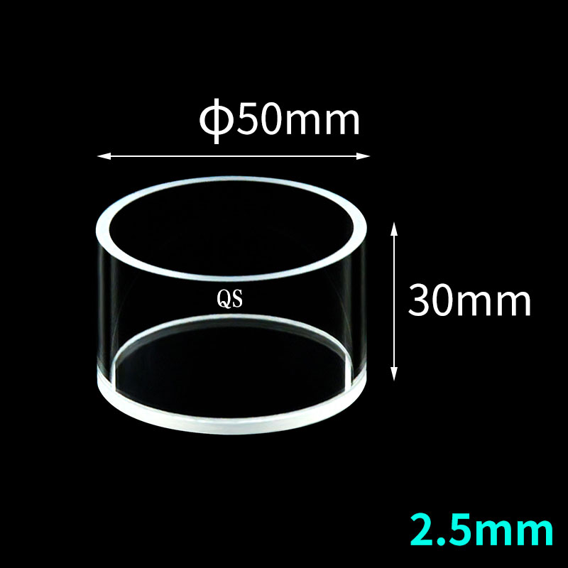
φ50 mm · C501WS1
For Avantes / smaller spheres
View C501WS1 →
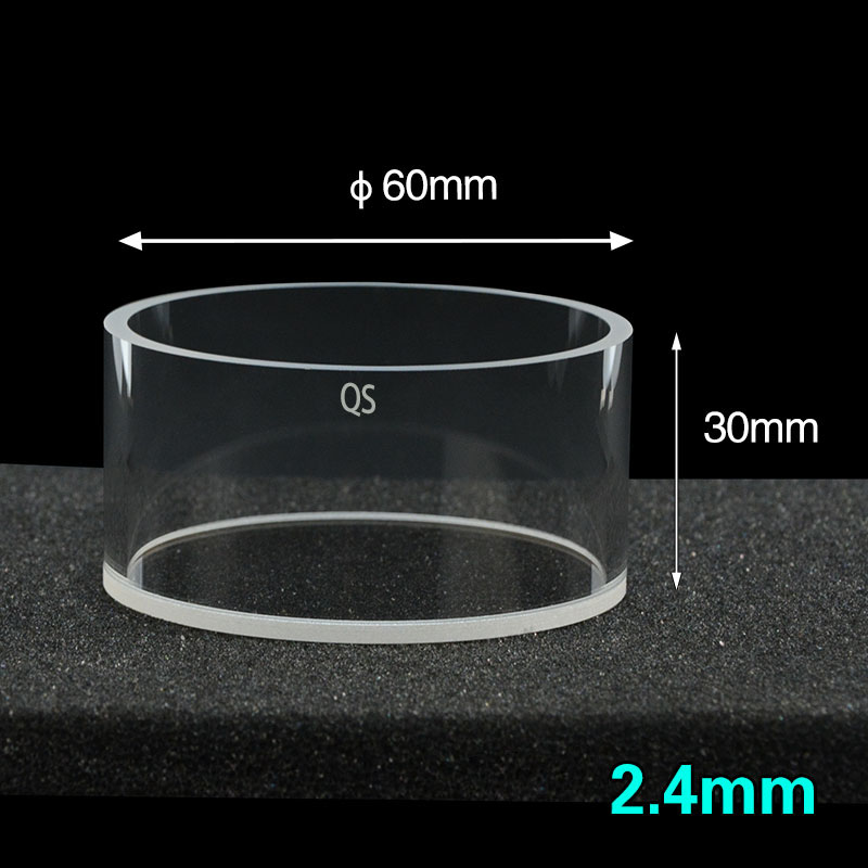
φ60 mm · C601WS ★
Cary / Lambda / Shimadzu standard
View C601WS →
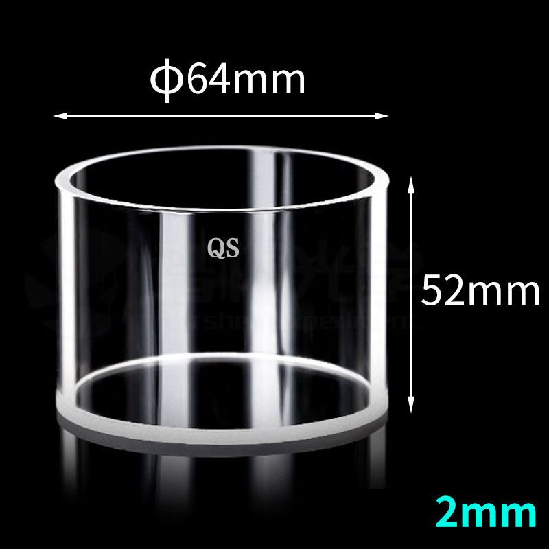
φ64 mm · C641WS1
Lambda 1050 with 150 mm sphere
View C641WS1 →
For PerkinElmer Lambda 950 / 1050 (150 mm sphere)
The Lambda 1050 with the L2D-7600 150 mm sphere accepts φ64 mm cuvettes directly, or φ60 mm cells with a port reducer. We stock both:
| SKU pattern | Outer ⌀ | Interior | Best for |
|---|---|---|---|
| C641WS1 / C641WS | φ64 mm | φ60 × 36–50 mm | Lambda 1050 native fit, deep samples |
| C601WS | φ60 mm | φ55.2 × 27.6 mm | Lambda 1050 with reducer port |
For Avantes / Ocean Optics small spheres
Compact benchtop spectrometers (Avantes AvaSpec / AvaSphere series, Ocean Optics ISS / FOIS) use 30–50 mm sphere ports. We stock the matching cylindrical cells:
| Sphere model | Port ⌀ | MQ cuvette |
|---|---|---|
| AvaSphere-50 | 50 mm | C501WS family |
| AvaSphere-30 / Ocean Optics ISS-30 | 30 mm | C301WS family |
| Avantes AvaSphere-15 / OEM compact | 12.5–17 mm | C12-51WS / C171WS family |
Custom diameters and special configurations
For diameters outside the standard 13 sizes (e.g., φ75 mm for non-standard pharmaceutical PAT, φ8–10 mm for OEM optical sensors, jacketed cells with temperature control, fully sealed cells for moisture-sensitive samples), MachinedQuartz quotes custom Sintered 83 cells with 4-week lead time. Submit a request via the custom quartz cuvettes form with your spectrometer model and sample type.
For the full SKU list with stock status, see the Quartz Cylinder Cuvettes catalog. For path-length-style analysis on the depth-vs-Kubelka-Munk question, the Beer-Lambert path length calculator can give a starting estimate by entering your sample’s apparent absorbance at the target wavelength.
Frequently asked questions
What is a cylindrical cuvette used for?
Diffuse reflectance UV-Vis spectroscopy on opaque samples — powders, slurries, films, plastic plates, and any solid you cannot see through. The cell sits at the entrance port of an integrating sphere accessory; sample is loaded into the chamber from the top; the optical face contacts the sphere interior. Cylindrical cuvettes are not used for transmission spectroscopy — that’s what rectangular cuvettes are for.
How is diffuse reflectance different from transmission UV-Vis?
Transmission passes the beam through a clear sample and follows Beer-Lambert law (A = ε·c·l). Diffuse reflectance illuminates an opaque or scattering sample and measures the total reflected intensity collected by an integrating sphere. The math uses Kubelka-Munk theory rather than Beer-Lambert. Cuvette geometry differs accordingly — rectangular for transmission, cylindrical for reflectance.
Do I need an integrating sphere to use a cylindrical cuvette?
Yes. Cylindrical reflectance cuvettes are designed to fit the sample port of an integrating sphere accessory. Without an integrating sphere, the cell has no useful optical function. If your spectrometer didn’t ship with an integrating sphere, you have to buy one as an accessory before any cylindrical cell becomes useful — typical accessory cost is $5,000–$25,000 depending on the spectrometer brand and sphere size.
How do I pick the right cylindrical cuvette diameter for my spectrometer?
Match the cuvette outer diameter to your integrating sphere’s sample port diameter — the spec is usually listed in the integrating sphere accessory’s manual under “sample port diameter”. Most modern instruments use 60 mm ports and need a φ60 mm cuvette. Premium high-end systems (Lambda 1050 with 150 mm sphere) take φ64 mm. Compact systems (Avantes, Ocean Optics, OEM) use smaller diameters from φ12.5 to φ50 mm.
How deep does the sample need to be in a cylindrical cuvette?
Deep enough to satisfy the Kubelka-Munk infinite-thickness criterion at your measurement wavelength. For strongly absorbing samples (dark powders, melanin), 3 mm is enough. For typical pharmaceutical APIs and polymers, 5–8 mm. For weakly absorbing white powders and lightly-pigmented samples, 15–25 mm. The safe rule is to order the deepest cell your sphere accommodates and fill it; excess depth never hurts.
Why are MachinedQuartz cylindrical cuvettes only made in Sintered 83?
Diffuse reflectance involves thousands of light bounces inside the integrating sphere. Each bounce that touches a glue interface (in Standard 80 cells) picks up trace UV-active emission from the adhesive, producing a baseline shift of 0.5–2% in the 240–340 nm region. Sintered 83 cells are powder-fused into one piece with no adhesive anywhere, eliminating this artifact. For pharmaceutical QC and high-precision color measurement, Sintered 83 is the only fabrication that produces clean DRS data.
Can I use a cylindrical cuvette for liquid samples?
Yes, for opaque or scattering liquids — milk, latex, slurry, suspension. The cell holds the liquid in the cylindrical chamber and presents it to the integrating sphere. For clear liquids you should use a rectangular transmission cuvette instead — diffuse reflectance on a transparent liquid gives nothing useful because the light passes through to the back wall and bounces back at unpredictable angles.
How do I clean a cylindrical reflectance cuvette?
Empty the sample, rinse with sample-compatible solvent first, then submerge the whole cell in 1% Hellmanex at 50 °C for 30 minutes, sonicate at 40 kHz for 5 minutes while submerged, triple DI rinse, ethanol displacement rinse, then air-dry rim-up overnight. The cylindrical geometry traps detergent more than rectangular cells, so 5× rinse minimum. Full procedure in the cleaning protocol guide.
What sample preparation is required for diffuse reflectance?
Three things matter most: particle size (grind to consistent < 100 µm for routine work, < 50 µm for chemometric calibration), packing density (gently tap to a uniform density across all samples — same protocol every time), and surface flatness (level the top of the powder column with a flat-bottom plate before measurement). Variation in these three is the largest source of systematic error in DRS calibration.
Where can I find custom-diameter cylindrical cuvettes?
MachinedQuartz quotes custom Sintered 83 cylindrical cells in any outer diameter, interior diameter, and depth combination at 4-week lead time. Common custom requests: φ75 mm for non-standard PAT systems, φ8–10 mm for OEM optical sensors, jacketed cells with thermal control for in-situ DRS, sealed cells for moisture-sensitive pharma samples. Submit your spectrometer model and sample type via the custom quartz cuvettes quote form.
About this guide. MachinedQuartz manufactures cylindrical reflectance cuvettes for diffuse-reflectance UV-Vis spectroscopy customers across pharmaceutical R&D, polymer characterization, color/cosmetics, catalyst research, and battery materials. Spectrometer compatibility, Kubelka-Munk depth thresholds, and sample-preparation guidance come from internal QC measurements and customer reference setups across 30+ standard SKUs.
Next step: confirm your sphere port, pick the cell
Picking the right cylindrical reflectance cuvette comes down to three numbers: outer diameter (must match your spectrometer’s sphere port), interior dimensions (sample volume × depth for Kubelka-Munk), and fabrication (Sintered 83 default, Molded 83 for elevated-temperature in-situ work).
For non-standard configurations or custom outer diameters, MachinedQuartz quotes custom cells with 4-week lead time. Standard SKUs ship in 1–3 days from US stock.


Recent Comments