
Cuvette Path Length by Analyte: Concentration-Based Selection Matrix
This post is also available in:
Choosing cuvette path length by analyte concentration is the practical application of Beer-Lambert (A = ε·c·L) at the bench: keep the measured absorbance in the 0.1–1.0 AU linear range by selecting a path length that matches your sample. Use sub-mm cells (0.01–0.5 mm) for concentrated proteins and undiluted dyes; standard 10 mm for analytes at 1–100 µM; long-path 50–100 mm for trace analytes below 100 nM. The MQ path-length-by-analyte matrix maps 40+ common analytes to recommended path lengths.
Selection Reference · By Analyte Class
Cuvette Path Length by Analyte: Concentration-Based Selection Matrix
Pick the right cuvette path length for proteins, nucleic acids, fermentation broths, dyes, transition-metal colorimetric assays, petroleum products, and environmental water samples — with the concentration ranges, target absorbance values, and stock SKUs that get you measuring at the first attempt.
8
Analyte Classes
0.1–1.0
Optimal AU Window
0.01–100 mm
Path-Length Range
60+
Stock SKUs
Choosing a cuvette path length is the most consequential decision in routine UV-Vis spectrophotometry, and it should not be a guess. The Beer-Lambert law — A = ε · c · l — tells you that absorbance scales linearly with both concentration c and path length l. If you cannot change the molar absorptivity ε (it is a property of your analyte at the chosen wavelength) and you do not want to dilute your sample, the path length is the variable you are actually selecting.
This guide reduces the selection problem to a per-analyte answer. For eight analyte classes that cover most lab UV-Vis traffic — proteins, nucleic acids, enzyme cofactors, pigments and dyes, fermentation broths, transition-metal colorimetric chemistry, petroleum products, and environmental water samples — we list the typical concentration range you encounter, the target absorbance window, and the path length that lands you in the linear-detection sweet spot of any modern spectrophotometer. The page closes with a note on why fluorescence selection is different and a link to our Beer-Lambert path length calculator for cases where you want to plug in your specific numbers.
How to use this page. Jump to your analyte section if you already know what you are measuring. If you are setting up a new method, start at section 1 for the 0.1–1.0 AU rule, then read the per-analyte recommendations. Every recommendation maps to a specific stock SKU in the MachinedQuartz catalog so you can quote and order without translating “5 mm cuvette” into a part number.
1. The 0.1–1.0 AU rule — the sweet spot you are aiming for
Modern UV-Vis spectrophotometers are linear and reproducible across the absorbance range of approximately 0.1 to 1.0 AU. Above 1.0 AU the photometric noise increases (you are measuring a small difference between two small intensities); above 2.0 AU most instruments saturate and the detector cannot distinguish the transmitted intensity from background. Below 0.1 AU the relative noise on a low-signal measurement degrades precision (a 0.001 AU drift on a 0.05 AU peak is a 2 % error). The sweet spot is 0.2–0.8 AU; the workable range is 0.1–1.0 AU.
For any given sample, three things determine where you land on the absorbance scale:
- The molar absorptivity ε of the analyte at the chosen wavelength — a property of the molecule, not under your control.
- The concentration c of the analyte — under your control through dilution, but dilution adds error and consumes sample.
- The path length l of the cuvette — under your control by selecting a different cell.
If you do not want to dilute (precious sample, tedious dilution series, or you need to preserve the original concentration for downstream work), the path length is the only variable you are choosing. The remaining sections of this page give you that decision per analyte class.
The 2.0 AU saturation cliff. Above approximately 2.0 AU you are measuring 99% absorption versus 99.9% absorption — a 10x difference in transmitted intensity to a number that is essentially noise. The exact ceiling depends on the instrument (modern double-beam scanners reach 3.0 AU; diode-array spectrometers often saturate at 2.5 AU). Aim well below the cliff: above 1.5 AU, switch to a shorter path length.
 DNA / Protein
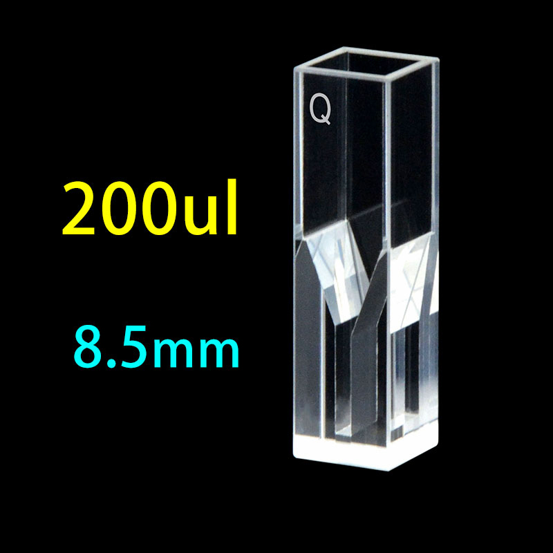
C104CS99 — 10 mm ultra-micro
200 µL · masked aperture · the workhorse for A260 nucleic acid & A280 protein
View micro cuvettes →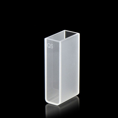 OD600 / Colorimetric
20 mm standard cuvette
7 mL · two-way light · for fermentation OD600, Bradford / Lowry / BCA, transition-metal colorimetric
View standard cuvettes →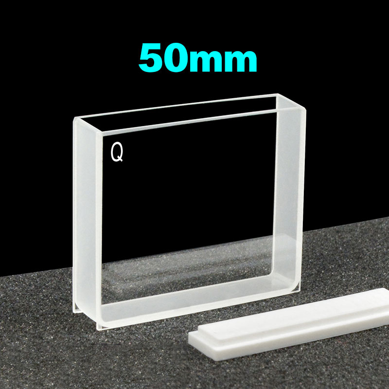 Trace water QC
C502CA5 — 50 mm long-path
17.5 mL · 5× sensitivity vs 10 mm · for trace nitrate / phosphate / chlorine / pharma impurity
Long-path guide →2. Quick lookup matrix
Find your analyte class. Read across to the recommended path length and the wavelength most analysts use. If your concentration falls at the edge of the typical range, refer to the dedicated section below for guidance on stepping up or down.
| Analyte class | Wavelength | Typical c | Recommended path | If concentrated | If trace |
|---|---|---|---|---|---|
| Proteins (A280) | 280 nm | 0.05–50 mg/mL | 1 mm | 0.1–0.5 mm | 10 mm |
| Bradford / Lowry / BCA | 595 / 750 / 562 nm | 0.1–1.5 mg/mL | 10 mm | 5 mm | 10 mm + concentrate |
| DNA / RNA | 260 nm | 10–3000 ng/µL | 1 mm | 0.1–0.5 mm | 10 mm micro |
| NADH / NADPH kinetics | 340 nm | 10 µM–1 mM | 10 mm | 5 mm | 50 mm or fluorescence |
| Pigments / dyes | 400–700 nm | 0.1–100 µM | 10 mm | 1–5 mm | 50–100 mm |
| Fermentation OD600 | 600 nm | 0.05–5.0 OD | 10 mm | 1–5 mm dilute | 10 mm + concentrate |
| Transition-metal colorimetric | 500–700 nm | 0.1–100 mg/L | 10 mm | 1 mm | 50 mm |
| Petroleum (API / Saybolt) | 520 / 530 nm | By color scale | 10 mm | N/A method-defined | N/A |
| Trace water (Cl/NO₃/PO₄) | 220–540 nm | 10 µg/L–100 mg/L | 50 mm | 10 mm | 100 mm |
The middle column (“Recommended path”) is the single best choice for typical concentrations. Use the right two columns when your sample sits at the edge of the typical range. Multi-path workflows (running 1, 5, and 10 mm cuvettes in parallel for the same sample) are covered in how to choose UV-Vis cuvette path length.
3. Proteins — A280, BCA, Bradford, Lowry
Protein quantitation has two distinct cases that need different path lengths.
Direct A280 absorbance
Aromatic amino acids (tryptophan, tyrosine, and to a lesser extent phenylalanine) absorb at 280 nm with an effective molar absorptivity around 1.0–1.5 (mg/mL)⁻¹ cm⁻¹ for a typical mixed protein. At 1 mg/mL in a 10 mm cuvette, A280 lands near 1.0 AU — right at the edge of the linear window.
- 0.05–0.5 mg/mL: use a 10 mm cell.
- 0.5–5 mg/mL: use a 1 mm cell — the workhorse choice for most lab proteins.
- 5–50 mg/mL (concentrated antibody or recombinant protein stock): use a 0.1 mm or 0.5 mm cell, or a demountable thin-film holder.
- Above 50 mg/mL: dilute, or measure in a 0.05 mm demountable cell.
Colorimetric assays (Bradford, Lowry, BCA)
Bradford reads at 595 nm (Coomassie Brilliant Blue G-250 dye binding), Lowry at 750 nm (alkaline copper-protein-Folin), and BCA at 562 nm (bicinchoninic acid copper reduction). All three are calibrated against BSA or IgG standard curves and target a working range of 0.1–1.5 mg/mL.
- Standard 10 mm cuvettes are the published method default and the right choice for almost all colorimetric protein work.
- For microtiter-plate formats (96-well), the effective path length is 5–6 mm at 200 µL well volume; calibration curves must be re-done because the path length differs from cuvette work.
SKU recommendation
Standard 10 mm A280 work
Catalog 10×10×40 mm two-clear-side cuvette, JGS1 quartz for 280 nm UV transmission. See size chart.
SKU recommendation
Concentrated protein 1–0.5 mm
Sub-micro 1 mm or 0.5 mm path quartz cuvette — 50–200 µL sample volume. Micro cuvette guide.
4. Nucleic acids — DNA and RNA at A260
Nucleic acids absorb at 260 nm with an effective extinction coefficient of approximately 50 (µg/mL)⁻¹ cm⁻¹ for double-stranded DNA, 40 for single-stranded DNA, and 33 for RNA. At 50 ng/µL of dsDNA in a 10 mm cuvette, A260 = 1.0 AU — saturating the linear window for most genomic-prep concentrations.
- Diluted DNA < 20 ng/µL: use a 10 mm cell. Common for sequencing prep dilutions.
- Standard genomic prep, 50–500 ng/µL: use a 1 mm cell. This is where most molecular biology lives.
- Concentrated stocks, 500–3,000 ng/µL: use a 0.1 mm or 0.5 mm cell.
- Plasmid prep over 3,000 ng/µL: dilute first, then read at 1 mm; or use a microvolume pedestal (see cuvette vs NanoDrop comparison, coming soon).
Purity ratios A260/A280 and A260/A230
Purity ratios are independent of path length — both numerator and denominator scale with l — so any well-chosen path length gives the same ratio. The exception: at very low absorbance (< 0.1 AU at either wavelength) the ratio becomes unreliable because instrument noise dominates the lower-absorbance reading. If your A260 is below 0.1, switch to a longer cuvette so the ratio calculation has signal to work with.
Sample volume. A 1 mm sub-micro cuvette typically holds 100–200 µL; a 0.5 mm holds 50–100 µL. Both are excellent for DNA work where sample is precious. For volumes below 50 µL, consider a 0.5 mm demountable cell or a microvolume pedestal.
5. Enzyme & cofactor assays — NADH, NADPH, NAD⁺, NADP⁺
NADH and NADPH absorb at 340 nm with an extinction coefficient of 6,220 M⁻¹ cm⁻¹. At 100 µM NADH in a 10 mm cuvette, A340 ≈ 0.62 AU — comfortably mid-window. NAD⁺ and NADP⁺ (oxidised forms) do not absorb at 340 nm; the appearance and disappearance of the 340 nm band is the readout for most NAD-dependent dehydrogenase assays.
- Standard kinetic assays at 10–500 µM NADH: use a 10 mm cell. The default for malate, lactate, glutamate, alcohol dehydrogenase, glucose-6-phosphate dehydrogenase, and most clinical chemistry methods.
- Trace-level kinetics < 5 µM NADH: use a 50 mm cell, or switch to fluorescence detection (NADH fluoresces at 460 nm with 340 nm excitation, with much higher sensitivity than absorbance).
- Above 1 mM NADH: dilute first, or use a 1–5 mm cell.
For temperature-controlled enzyme kinetics, a screw-top sealed cuvette reduces evaporation during 30+ minute kinetic runs and pairs cleanly with thermostatted Peltier holders.
6. Pigments & dyes — visible-range chromophores
Natural and synthetic chromophores cover a wide range of extinction coefficients. Chlorophyll a at 664 nm has ε ≈ 76,000 M⁻¹ cm⁻¹ (very strong); methyl orange at 464 nm has ε ≈ 27,000 M⁻¹ cm⁻¹; food dyes are often around 10,000–30,000 M⁻¹ cm⁻¹.
- Standard 10 mm for analytical work in the 0.1–100 µM range — covers most dye chemistry, food colour, beverage colour, and chlorophyll extracts after dilution.
- 1–5 mm for concentrated stock dye solutions, or for in-process colour QC where the working solution is the production concentration.
- 50 mm for trace dye contamination work (residual dye in wastewater, decolourisation efficiency, environmental dye traces).
Visible vs UV cuvettes
Pigment and dye work is overwhelmingly in the visible (400–700 nm) where standard JGS2 quartz, optical glass, and even disposable plastic cuvettes are all transparent. JGS1 deep-UV grade is unnecessary unless your method also reads below 220 nm. See our UV cutoff guide for the trade-off.
7. Fermentation & cell culture — OD600
Optical density at 600 nm is a turbidity measurement, not a true absorbance — it measures forward scatter from cells. Beer-Lambert linearity holds approximately up to OD600 ≈ 0.6–0.8 in a 10 mm cuvette, then deviates because of multiple-scattering. For comparable historical numbers, the convention is to dilute concentrated cultures back into the linear window of a 10 mm cell.
- 10 mm cuvette is the published method default for E. coli, yeast, mammalian cell suspensions, and most fermentation work. All published growth curves and OD-vs-CFU calibrations assume 10 mm path length.
- For dense cultures > OD 1.0, dilute 1:5 or 1:10 in growth medium and read in a 10 mm cuvette. Avoid the temptation to switch to a 1 mm cell for dense cultures — you will get a number, but it will not match published growth curves and the multiple-scattering bias scales with the cell concentration in non-trivial ways.
- For continuous online monitoring, in-line transmission probes with a 5 mm or 10 mm effective path length give real-time OD without sampling.
Different spectrophotometers give different OD readings. The numerical value of OD600 depends on the spectrometer geometry (slit width, detector aperture, distance from cell to detector). Always validate a new instrument against your existing growth curves before changing the workflow. Path length is one variable; instrument geometry is another.
8. Transition-metal colorimetric chemistry
The classical wet-chemistry methods — permanganate, dichromate, iron with phenanthroline, copper with neocuproine, manganese with periodate, ammonia with Nessler — all develop coloured complexes with extinction coefficients in the 4,000–30,000 M⁻¹ cm⁻¹ range and target the 500–700 nm visible window.
- Standard 10 mm cuvette covers 0.1–100 mg/L of analyte for most published methods. Standard Methods, ASTM, and EPA protocols all assume 10 mm path length unless explicitly stated otherwise.
- 1 mm cuvette for concentrated process samples (electroplating bath QC, high-strength industrial wastewater).
- 50 mm cuvette for trace metals at the µg/L level — commonly used in environmental water analysis and ultrapure-water QC where the regulatory limit is well below 1 mg/L.
Colour development time
Many transition-metal methods require fixed colour-development time (5, 15, 30 minutes depending on the method). Read all standards and samples at the same elapsed time after reagent addition. Path length does not change the kinetics of colour development; it just determines what concentration band lands in your absorbance window.
9. Petroleum products — API color, Saybolt, ASTM color
Petroleum colour is a regulatory and customer-facing specification, not a quantitative absorbance number. Three method standards dominate:
- ASTM D1500 (520 nm): dark petroleum products (lubricating oils, fuel oils, residual fuels). Uses a 33-mm path-length cell against an octane standard.
- ASTM D156 / Saybolt (540 nm): light products (kerosene, naphtha, white oils). Uses a 50-mm or 100-mm path-length cell.
- ASTM D6045 (525 nm): spectrophotometric automation of D1500 and D156 in a 10-mm cell, used by colorimeters in refineries.
Path length here is method-defined, not chosen by the analyst. Use the cell length that matches the method standard or your colorimeter manufacturer’s calibration. The relevant question is whether the cuvette material (path length aside) is correct: ASTM petroleum methods accept optical glass and quartz interchangeably for the colour wavelengths used.
10. Environmental water analysis — trace UV-Vis
Trace inorganic and organic analytes in water sit in the µg/L to mg/L range, well below the typical concentrations of fermentation or pharmaceutical work. The published EPA and Standard Methods protocols use long-path cuvettes for two reasons: low concentration and low molar absorptivity for many of the relevant analytes.
- Free chlorine (DPD method, 530 nm): 10 mm cell at 0.05–5 mg/L; 50 mm cell at 0.005–0.5 mg/L. The 100 mm cell takes the working range down to single-µg/L for ultrapure-water QC.
- Nitrate (UV 220 nm or cadmium reduction 540 nm): 50 mm cell standard for drinking water (0.1–10 mg/L NO₃-N); 100 mm for source-water trace.
- Phosphate (molybdenum-blue, 880 nm): 10 mm cell at 0.1–10 mg/L; 50 mm cell at 10 µg/L–1 mg/L; 100 mm for environmental trace.
- Nitrite (Griess reaction, 540 nm): 10 mm cell standard at 0.01–1 mg/L; 50 mm for trace.
- Iron (phenanthroline, 510 nm): 10 mm at 0.1–5 mg/L.
The 50 mm and 100 mm cuvettes used for trace water work are a major commercial niche of their own — covered in our upcoming long-path cuvettes for trace UV-Vis analysis guide. They use the same JGS-grade quartz and the same fabrication discipline as our standard 10 mm cells, just with a longer outer dimension.
11. Fluorescence is a different problem
The path length analysis above applies to absorbance spectroscopy. For fluorescence, the geometry changes the analysis:
- The excitation path traverses the cuvette once on its way in. Path length matters for excitation absorption, but only weakly — fluorometers typically use a fixed 10 mm cuvette and accept whatever excitation path length that is.
- The emission collection path at 90 degrees to excitation is what limits sensitivity. Geometry of the cell (square 10x10x10 mm with all four sides polished, or short-aperture for low-volume samples) matters more than path length per se.
- Inner-filter effects — reabsorption of emitted fluorescence by the analyte itself — become significant when absorbance at the excitation wavelength exceeds about 0.05 AU in a 10 mm cell. Above this concentration, fluorescence intensity is no longer linear in concentration. The fix is to dilute (preserves cell choice) or use a shorter path-length fluorescence cell.
For a complete treatment of fluorescence cuvette geometry, polish requirements, and inner-filter management, see our fluorescence cuvette guide.
12. Tools and stock SKUs by path length
For numerical work, plug your specific concentration, extinction coefficient, and target absorbance into the calculator and let it return the right path length. For procurement, jump to the size chart and select by outer dimensions.
Need a custom path length?
Standard cells from 0.01 mm to 100 mm ship from stock. Custom path lengths within that range carry no tooling fee on geometry that fits our standard envelope.
Request custom quote →See bulk programs →13. Frequently asked questions
What path length should I use for protein A280?
For typical proteins at 0.5 to 5 mg/mL, use a 1 mm cuvette. For dilute proteins below 0.5 mg/mL, use a 10 mm cell. For concentrated stocks above 5 mg/mL, use a 0.1 to 0.5 mm cuvette or a demountable thin-film holder. The single most-common pick for routine A280 work is a 1 mm sub-micro cuvette holding 100 to 200 microliters.
What is the optimal absorbance window for UV-Vis?
0.1 to 1.0 absorbance units (AU) is the optimal window for modern UV-Vis spectrophotometers, with 0.2 to 0.8 AU being the sweet spot. Above 1.0 AU photometric noise increases; above 2.0 AU most instruments saturate. Below 0.1 AU relative noise on a low-signal measurement degrades precision. The path length you choose should land your analyte in this window at its typical concentration.
Can I use a 1 mm cuvette for fermentation OD600?
It will give you a number, but the number will not match published growth curves. OD600 protocols and CFU calibrations universally assume a 10 mm path length, and the multiple-scattering bias from cell density does not scale linearly with path length. Always dilute dense cultures (OD over 1.0) into the linear window of a 10 mm cell rather than switching to a shorter path.
What path length do I need for trace water analysis?
For drinking-water quality (chlorine, nitrate, phosphate at sub-mg/L levels), a 50 mm cuvette is the standard choice and lets you reach the linear AU window with the typical analyte concentrations. For ultrapure-water QC at single-microgram-per-litre concentrations, step up to a 100 mm cell. Both sizes are stocked in JGS quartz; see our upcoming long-path cuvettes guide.
Does path length affect the A260/A280 purity ratio?
No. Both A260 and A280 scale with the same path length, so the ratio is independent of cuvette choice. The only practical caveat is that at very low absorbance (below 0.1 AU at either wavelength), instrument noise dominates and the ratio becomes unreliable. If your A260 is below 0.1, use a longer cuvette so the calculation has signal to work with.
How do I read concentrated DNA above 1000 ng/uL?
Use a 0.1 mm or 0.5 mm cuvette. At 1000 ng/microlitre of double-stranded DNA in a 1 mm cell, A260 is around 2.0 AU (saturating most instruments). Switching to 0.1 mm drops the path length tenfold and brings you back into the linear window. Sub-millimetre cells require careful filling (no bubbles, no meniscus distortion) but give linear, reproducible readings on concentrated samples.
Why do I get different OD600 numbers on different instruments?
OD600 depends on instrument geometry: slit width, detector aperture, sample-to-detector distance, and stray light all affect the apparent reading. Path length is one variable, but two spectrometers with different geometry will give different OD readings on the same sample even at the same path length. Always validate a new instrument against your existing growth curves before you switch workflows.
Is JGS1 quartz necessary for visible-range pigment work?
No. JGS1 deep-UV grade is needed only when your method reads below 220 nm. For visible work (400 to 700 nm) including pigment, dye, and most colorimetric chemistry, standard JGS2 quartz, optical glass, and even disposable plastic cuvettes are equally transparent. JGS1 costs more without delivering benefit in the visible range. See our UV cutoff guide for the wavelength-by-grade trade-off.
Why does the petroleum industry use a 33 mm cell?
It is a method-defined choice in ASTM D1500 (color of petroleum products). The 33 mm cell length, combined with a tungsten lamp and 520 nm wavelength, was calibrated against the historical Lovibond color scale used by refineries. Modern colorimeters can substitute a 10 mm cell with conversion (ASTM D6045), but the 33 mm cell is still the reference standard for arbitration. Choose the cell that matches your method standard.
How do I select a path length when I do not know my analyte concentration?
Start with a 10 mm cuvette and read your sample. If absorbance is above 1.5 AU, dilute or switch to a shorter path. If absorbance is below 0.05 AU, switch to a longer path. With one or two iterations you will land in the 0.1 to 1.0 AU window. Once you know the typical concentration of your sample, the per-analyte sections above tell you the right starting cuvette for the next batch.
14. Disclaimer & notes
Path-length recommendations on this page are general guidance based on typical analyte concentrations and the 0.1–1.0 AU detection window of modern UV-Vis spectrophotometers. Your specific assay, sample matrix, instrument geometry, and method standard may require different choices. For pharmacopoeial methods (USP, EP, JP) and regulatory protocols (EPA, ASTM), follow the path length specified in the method.
Method-defined path lengths — petroleum colour (ASTM D1500: 33 mm), Saybolt (D156: 50 or 100 mm), DNA quantitation guidance (variable by lab SOP), and pharmaceutical assay methods — supersede any general guidance on this page. When in doubt, follow the method.
Concentration ranges are typical for the listed analyte class. Specific samples may sit outside the typical range; multi-path workflows (running 1, 5, and 10 mm cells in parallel) cover the edge cases when sample variability is high.
Stock SKU references are based on our current catalog. SKU availability and lead times may change. For binding pricing and lead times, see the bulk & OEM page or contact us directly.
Information currency: last reviewed May 2026.
Further reading


Recent Comments