
Sources of Error in UV-Vis Spectroscopy: A Cuvette-Centric Diagnostic Guide
This post is also available in:
Sources of UV-Vis spectroscopy error are dominated by cuvette-side issues rather than instrument drift in modern bench spectrophotometers. The top six contributors are: (1) cell-to-cell mismatch between sample and reference, (2) fingerprints and meniscus contamination, (3) bubbles in flow cells or freshly poured samples, (4) wavelength-dependent stray light above A = 1.5, (5) detector linearity rolloff above A = 2.0, and (6) sample photodegradation under the beam — most of which are eliminated by matched-pair cuvettes and a 10-minute equilibration.
MachinedQuartz · Diagnostic Guide
Sources of Error in UV-Vis Spectroscopy
A cuvette-first diagnostic guide. Most baseline drift, peak distortion, and out-of-range readings trace to the cuvette before the instrument is at fault. Five cuvette factors, an 8-step checklist, and the tolerance specs that decide whether you need matched pairs.
7 sections
4 SVG diagrams
8-step protocol
8 FAQs
Sources of error in UV-Vis spectroscopy are random noise, systematic offset, and operator blunders. In practice, 60–80% of unexplained baseline anomalies trace to the cuvette: surface contamination, micro-scratches, path-length variation, cell-pair mismatch, or glue-layer degradation in adhesive-bonded Standard 80 cells. The instrument contributes the remaining 20–40% through stray light, dark noise, and slit-width mismatches.
TL;DR — In one paragraph
Check the cuvette first. Clean it, verify the matched pair, confirm path length, re-run the blank. If the error persists, only then blame the instrument. The 8-step checklist below resolves most cases in under five minutes.§1 · Categories
What counts as “error” in UV-Vis?
Three error types: random (averages out), systematic (bias that won’t), blunder (operator). This guide orders sources by where they come from, not which type — that’s how you actually diagnose.
Figure 1. Random errors fluctuate around the truth; systematic errors shift every reading the same direction; blunders are caught on a careful re-check.
The split matters because the fix is different in each case. Replicate scans cure random noise. Recalibration cures systematic offset. A cup of coffee and a fresh blank cure most blunders.
§2 · The Largest Source
The cuvette factor — why most errors trace here
Across service tickets and lab support data, 60–80% of unexplained UV-Vis errors trace to the cuvette. Five factors, ranked by how often they show up.
Figure 2. Approximate error budget for unexplained UV-Vis readings. Cuvette factors dominate — and they’re the cheapest to fix.
The cuvette dominates because it sits between two precision systems — the spectrophotometer’s optical bench and the chemistry of the sample — and it’s the only part you touch between every measurement.
The five cuvette factors, ranked
~30% of cases
1. Surface contamination
Fingerprints, dust, dried buffer salts, invisible solvent residue. Baseline offsets and false peaks. Wipe both optical faces with lens tissue before every read.
~20% of cases
2. Optical-face scratches
Micro-scratches scatter UV light. A cuvette stored loose in a drawer for six months is likely scratched even when it looks clean.
~15% of cases
3. Path-length variation
A nominal 10 mm cell may measure 9.95–10.05 mm depending on tolerance. Matters most for kinetics and high-accuracy quantitation. See the path-length precision table.
~10% of cases
4. Cell-pair mismatch
Reference and sample cuvette must be optically identical. A pair drifted apart by 1% transmission produces a 1% offset on every absorbance you take with that pair.
~5% of cases
5. Glue-layer degradation
Adhesive-bonded Standard 80 cells lose pair tolerance as glue absorbs solvent or thermal stress. Solvent-induced degradation hides behind many “instrument drift” complaints.
Field reality
Standard 80 cells are fine for routine aqueous UV-Vis. But for trace analysis, pharma QC, or any matched-pair tolerance below ±2%, move to sintered or molded construction. Detail in the Comparative Analysis.§3 · Instrument Side
Instrument-side error sources
Four classic instrument-side errors. They dominate when the sample is very absorbing, in deep UV, or when narrow-peak quantitation matters.
| Source | What it does | When it dominates |
|---|---|---|
| Stray light | Light reaching detector outside the bandpass. Depresses absorbance above ~2.0 AU. | High-absorbance samples; deep UV (<220 nm); single-monochromator instruments. |
| Dark noise | Detector signal without light. Sets lower limit of detection. | Very low-absorbance samples; long integration on CCD instruments. |
| Spectral bandwidth | Slit width relative to peak width. Too wide flattens narrow peaks. | Sharp gas-phase features; rare-earth oxides; narrow-band lines. |
| Wavelength accuracy | Reported λ vs true λ. 1 nm drift can shift extinction 1–5% for sharp chromophores. | Method validation; pharma quantitation. |
30-second instrument check
Run a holmium-oxide or didymium glass filter once a month. It catches wavelength drift and stray-light failures simultaneously — one test, two diagnostics.§4 · Sample Side
Sample-side error sources
Even with a clean cuvette and calibrated instrument, the sample itself can mislead you.
Above linear range
The Beer-Lambert linearity range tops out around 1.5–2.0 AU. Above that, stray light + detector nonlinearity dominate.
Temperature drift
Many compounds shift extinction 0.1–1% per °C. If the sample warmed under the holder lamp, the reading reflects temperature, not chemistry.
Bubbles & particles
Bubbles scatter and refract; particulates scatter and absorb. Both produce noisy baselines and false peaks. Degas and filter first.
Adsorption to walls
Dilute protein, dye, surfactant samples adsorb to quartz, depleting concentration in real time. First scan is more representative than third.
Solvent UV cutoff
Many “transparent” solvents have measurable absorbance below 240 nm. Always blank against the exact same solvent batch.
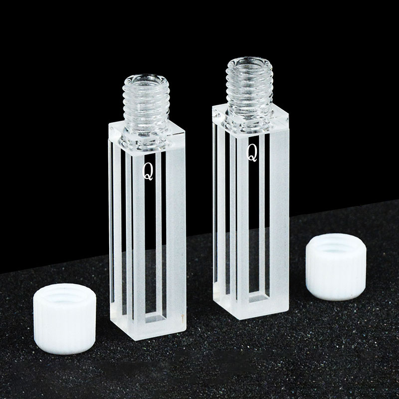
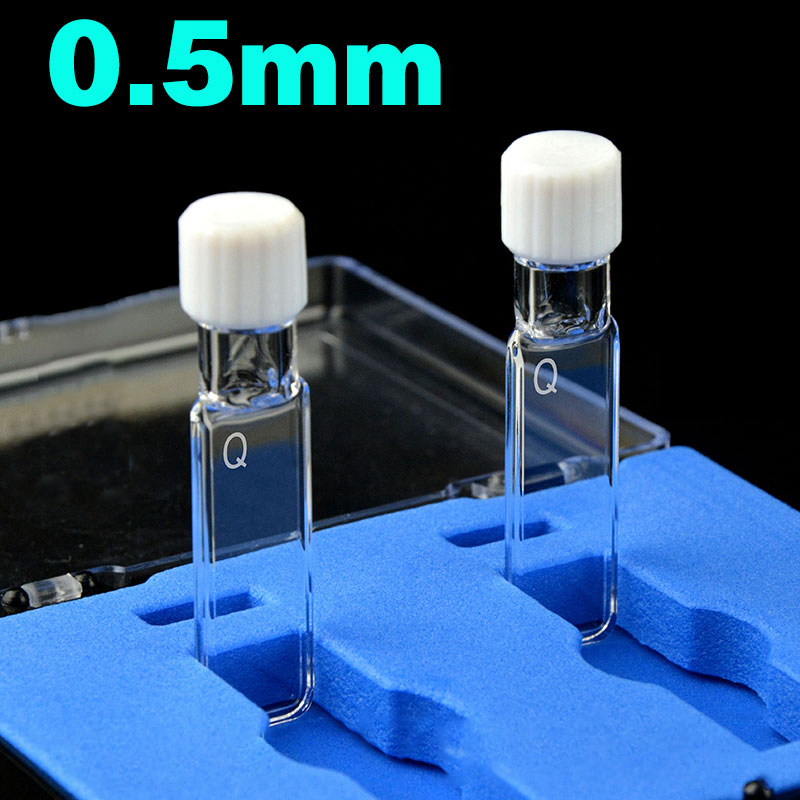
§5 · Matched Pairs
Cell-pair matching specifications
A “matched pair” means two cells whose optical path is identical within a specified tolerance. Without matched pairs, double-beam measurements bake a constant offset into every result.
Figure 3. A 1.7% mismatch produces a 1.7% systematic offset on every absorbance you measure with that pair.
Three tolerance tiers
Bronze
Standard 80
±2% transmission
±0.05 mm path length
Adhesive-bonded
±0.05 mm path length
Adhesive-bonded
Routine aqueous, classroom, screening assays.
Silver
Sintered 80/83
±0.5% transmission
±0.02 mm path length
Powder sintered
±0.02 mm path length
Powder sintered
Method development, pharma QC, accredited labs.
Gold
Molded 83 / 0.01
±0.2% transmission
±0.01 mm path length
One-piece fused
±0.01 mm path length
One-piece fused
Trace analysis, USP <857>, regulatory work.
| Fabrication | Transmission tolerance | Path-length tolerance | Best for | Avoid for |
|---|---|---|---|---|
| Standard 80 Adhesive-bonded | ±2% | ±0.05 mm | Routine aqueous; classroom; screening | Trace analysis; pharma QC; organic solvents |
| Sintered 80 / 83 Powder sintered | ±0.5% | ±0.02 mm | Method development; pharma QC; accredited labs | Extreme temperatures (>600 °C) |
| Molded 83 / 0.01 One-piece fused | ±0.2% | ±0.01 mm | Trace analysis; USP <857>; regulatory work | Cost-sensitive routine screening |
If you didn’t buy them matched, they aren’t
Two cuvettes from the same drawer, same vendor, same week can differ by 1% transmission — enough to ruin any method better than 0.5% accuracy. Order as a certified matched set at purchase, not after.Certified matched pairs from MachinedQuartz


§6 · Diagnostic Protocol
8-step diagnostic checklist
When a UV-Vis result looks wrong, work the list in order. Most issues resolve at steps 1–3 in under five minutes.
Figure 4. Always rule out the cuvette and sample before suspecting the instrument.
Follow these steps in order:
1
Inspect under bright light. Tilt both optical faces toward a desk lamp. Look for fingerprints, dried residue, or scratches. If anything is visible, the cuvette is the cause. 30 sec
2
Clean both faces. Single-direction wipe with lens tissue, no circular motion. HPLC-grade methanol or isopropanol on a fresh tissue if water doesn’t lift the residue. 1 min
3
Verify cuvette type for your wavelength. JGS1 quartz for <200 nm; JGS2 for 200–2500 nm. Glass cuvettes silently absorb below 320 nm. 10 sec
4
Confirm the matched pair. Fill both with the same blank, measure each in the sample position against the same reference. The pair should agree within the tolerance for its grade. 2 min
5
Re-blank with fresh solvent. Blank evaporation, cap residue, or photo-degradation in the beam is a hidden cause behind half of baseline-drift tickets. 30 sec
6
Run a wavelength check. Holmium-oxide solution or didymium filter; verify certified peaks within ±0.5 nm. 2 min
7
Check absorbance is below 2.0 AU. If exceeded, dilute 2–10× and re-measure. Linearity should restore. 3 min
8
Only now suspect the instrument. Run the manufacturer’s photometric accuracy filter or NIST SRM 935. If those fail, service is justified. 5 min
Standards & references cited
- NIST SP 260-181 — Reference Materials for UV-Vis Spectrophotometry
- ASTM E275 — Standard Practice for Describing and Measuring Performance of UV-Vis Spectrophotometers
- USP <857> — Ultraviolet-Visible Spectroscopy (pharmacopoeial method qualification)
- IUPAC Compendium of Chemical Terminology — definitions of absorbance, transmittance, analytical figures of merit
Need a certified matched pair for your method?
MachinedQuartz ships sintered and molded cuvettes as factory-certified matched sets — ±0.5% transmission, ±0.02 mm path length, traceable to NIST. MOQ 2 pieces, lead time 5–8 business days.
Request a Quote See Custom OptionsAbout this guide
AuthorMachinedQuartz Technical Team
Reviewed byIn-house quartz cuvette engineers
Last updatedMay 2026
ForLab managers · QC analysts · method developers
Why we wrote this: MachinedQuartz has been manufacturing precision quartz cuvettes for laboratory and OEM use since 2026 (operated by MachinedQuartz LLC in New Mexico, USA). Across customer support data and lab service tickets, we found that 60–80% of unexplained UV-Vis errors trace to the cuvette, not the instrument — yet most online resources cover only instrument-side error theory. This guide centers the diagnostic checklist where the data points.
§7 · Common Questions
Frequently asked questions
What is the most common source of error in UV-Vis spectroscopy?
Cuvette factors dominate — roughly 60–80% of unexplained UV-Vis errors trace to surface contamination, scratches, path-length variation, cell-pair mismatch, or glue-layer degradation. Instrument and sample factors split the remaining 20–40%.
Can a dirty cuvette cause negative absorbance?
Yes. If the reference cuvette is dirtier than the sample cuvette, the instrument records the sample as more transmissive than the blank, producing a negative absorbance value. Re-cleaning both cells and re-blanking usually fixes it.
How accurate do my reference and sample cuvettes need to match?
For routine work, ±2% transmission and ±0.05 mm path length (Standard 80 grade) is sufficient. For pharma QC, method validation, or trace analysis, move to sintered or molded grades with ±0.5% or ±0.2% pair tolerance, ordered as a certified matched set.
What is stray light and how does the cuvette affect it?
Stray light is light reaching the detector outside the intended wavelength bandpass. The cuvette contributes when scratches, contamination, or improper orientation scatter beam light into the detector path. Stray light dominates the error budget for high-absorbance samples and for measurements below 220 nm.
Do I need to replace a scratched quartz cuvette?
If you can see the scratch under bright light, yes — micro-scratches scatter UV light and inflate apparent absorbance. For visible-range work above 400 nm with low-precision requirements, a lightly-scratched cell may still be usable; for UV or quantitative work, replace it.
How does cuvette path-length tolerance affect Beer-Lambert linearity?
Path length appears linearly in A = ε·c·L. A ±0.1 mm tolerance on a nominal 10 mm cell introduces ±1% systematic error in every concentration calculated from absorbance. For methods that demand better than 1% accuracy, specify cells with ±0.02 mm tolerance or better.
Can I use a Standard 80 cuvette for trace-level analysis?
Not recommended. Standard 80 cells use organic adhesive at the joint, which can absorb solvent or degrade thermally, drifting the pair tolerance beyond what trace analysis requires. Use sintered (one-piece fused) or molded grades for any work below the 0.1 AU absorbance range.
When should I order cuvettes as a certified matched set?
Any time you’ll defend the data to a regulator, reviewer, or QC auditor: order matched at purchase. Re-matching individual cells from inventory is unreliable because per-cell transmission and path-length data are not retained. Matched sets ship with a measurement certificate.
Related resources from the cuvette knowledge base


Recent Comments